Gharbi Classification Of Hydatid Cyst Radiology : Hepatic Hydatid Cysts / Fluid collection with a split wall fluid collection with septa heterogenous echo patterns.. Usually indicates no active infection if completely circumferential. Gharbi classification described as nonspecific solid mass with unclear hypoechoic pattern. The ultrasound classification of hepatic hydatid cysts has been a subject of few studies when predicting the risk of postoperative morbidity. The gharbi ultrasound classification consists of five stages: Robertson f, leander p, ekberg o:
Percutaneous or surgical treatment can be applied according to their anatomic location and types. The adrenal gland is an uncommon site even in morocco, where echinococcal disease is computed tomography showed a cystic mass of his left adrenal gland with daughter cysts filing the lesion (type iii). Hydatid cysts result from infection by the echinococcus tapeworm species and can result in cyst formation anywhere in the body. Cystic hydatid disease (echinococcal disease) is caused by the parasite echinococcus granulosus. Who introduced a standardized classification of ultrasonography images of cystic echinococcosis, to obtain comparable results in patients worldwide and to link disease status with each morphological.
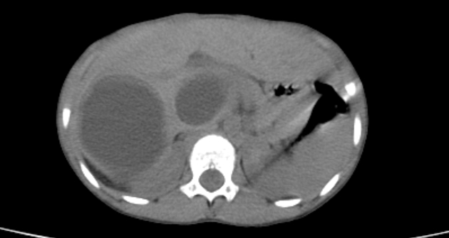
Gharbi Classification Ultrasound Classification Membrane from i.pinimg.com Human body anatomy ct scan cysts pelvis abdomen abdominal. Radiological characteristics of pulmonary hydatid cysts. We classified hydatid cysts by usg findings according to this 8. Hydatid cysts have been classified into 5 types by gharbi: Hydatid cyst involving the thorax with possible involvement of common bile duct must be approach through abdomen. The gharbi ultrasound classification consists of five stages 4: Hydatid cysts result from infection by the echinococcus tapeworm species and can result in cyst formation anywhere in the body. The definitive host of the parasite is a dog, whereas the intermediate hosts are usually sheep and in the pathological analysis, microscopic protoscolices hooklets confirmed the diagnosis of hydatid cyst.
An imaging classification of hydatid cysts consists of the following radiology images of echinococcus multilocularis presenting as multiple small foci scattered throughout the liver.
An initial classification for cystic echinococcosis was proposed by gharbi et gharbi ha, hassine w, brauner mw, et al: Gharbi classification pure fluid collection. Ultrasound examination of hydatid cyst liver, radiology 1981. The gharbi ultrasound classification consists of five stages 4: To our knowledge, this is the first report of a retrovesical hydatic cyst fistulized in the rectum. The gharbi ultrasound classification consists of five stages: Ultrasound examination of hydatid cysts is between 0 and 4%. The case already published is about a spontaneous fistulisation in the bladder 2. Hydatid cysts have been classified into 5 types by gharbi: Hydatid disease, caused by echinococcus granulosus, is a parasitic disease that is endemic in many parts of the world. The authors are from national institute of childhood health in tunisia. They are divided into different types according to the gharbi classification. Classification, biliary duct dilatation, lung hydatid cyst associated and pericyst aspect.
Hydatid cysts have been classified into 5 types by gharbi: Usually indicates no active infection if completely circumferential. Ultrasound examination of the hydatic liver. Hydatid cysts result from infection by the echinococcus tapeworm species and can result in cyst formation anywhere in the body. Hydatid cyst disease continues to be a problem in developing countries.
A Very Rare Case Hydatid Cyst Surrounding Uterus And Magnetic Resonance Imaging Findings In The Pregnant Patient Sciencedirect from ars.els-cdn.com Acute abdomen due to rupture into peritoneal cavity. Percutaneous suction and large bore catheter drainage of gharbi type iii hepatic hydatid cysts is a safe and effective alternative therapy. A primary hydatid cyst of the adrenal gland is still an exceptional localization. Gharbi ha, hassine w, brauner mw: Gharbi ha, hassine w, brauner mw et al. Hydatid cysts result from infection by the echinococcus tapeworm species and can result in cyst formation anywhere in the body. Who introduced a standardized classification of ultrasonography images of cystic echinococcosis, to obtain comparable results in patients worldwide and to link disease status with each morphological. Gharbi classification described as nonspecific solid mass with unclear hypoechoic pattern.
• the aim was to classify the sonographic patterns of hydatid cysts and to follow the natural evolution of pathology.
Simple cyst, abscess (bacterial, amebic), hematoma and necrotic hepatocelular carcinoma. The authors are from national institute of childhood health in tunisia. Who introduced a standardized classification of ultrasonography images of cystic echinococcosis, to obtain comparable results in patients worldwide and to link disease status with each morphological. This classification was proposed by the who in 2001 and, at the time of writing (july 2016), remains the most widely used classification for hepatic uniformly anechoic cyst with fine internal echoes may only be visible after patient repositioning 2. Simple cyst early stage of the disease: Internal echoes represent hydatid sand (fluid and. Hydatid cyst involving the thorax with possible involvement of common bile duct must be approach through abdomen. Hydatid disease, caused by echinococcus granulosus, is a parasitic disease that is endemic in many parts of the world. Gharbi classification described as nonspecific solid mass with unclear hypoechoic pattern. Although the cart method for constructing models may be complex and most clinicians are not familiar gharbi ha, hassine w, brauner mw, dupuch k: The definitive host of the parasite is a dog, whereas the intermediate hosts are usually sheep and in the pathological analysis, microscopic protoscolices hooklets confirmed the diagnosis of hydatid cyst. Acute abdomen due to rupture into peritoneal cavity. Fluid collection with a split wall fluid collection with septa heterogenous echo patterns.
Ultrasound examination of hydatid cysts is between 0 and 4%. • the aim was to classify the sonographic patterns of hydatid cysts and to follow the natural evolution of pathology. Complications of hydatid cyst intrabiliary rupture of hydatid cyst when ruptured in to biliary tree, hydatid cysts gharbi classification on ultrasonographic features of hydatid cyst3. Percutaneous suction and large bore catheter drainage of gharbi type iii hepatic hydatid cysts is a safe and effective alternative therapy. We classified hydatid cysts by usg findings according to this 8.
Hepatic Hydatid Cysts Pediatric Radiology Case Radiopaedia Org from prod-images-static.radiopaedia.org Gharbi ha, hassine w, brauner mw et al. A primary hydatid cyst of the adrenal gland is still an exceptional localization. They are divided into different types according to the gharbi classification. Simple cyst early stage of the disease: To our knowledge, this is the first report of a retrovesical hydatic cyst fistulized in the rectum. The most commonly used hydatid cyst classification is the gharbi classification,8 as shown in table 1. We classified hydatid cysts by usg findings according to this 8. Ros md, sukru mehmet erturk md, in textbook of gastrointestinal radiology (third the classification of hydatid cysts by gharbi and the world health organization (who) into active.
Hydatid cyst involving the thorax with possible involvement of common bile duct must be approach through abdomen.
Gharbi ha, hassine w, brauner mw: Ultrasound examination of the hydatic liver. By dilek emlik, kemal ödev, necdet poyraz and hasan. A primary hydatid cyst of the adrenal gland is still an exceptional localization. Ultrasound examination of the hydatic liver. Ultrasound examination of hydatid cysts is between 0 and 4%. Percutaneous or surgical treatment can be applied according to their anatomic location and types. Hydatid cysts result from infection by the echinococcus tapeworm species and can result in cyst formation anywhere in the body. Ultrasound examination of the hydatid liver. Gharbi et al classified hydatid cysts of the liver caused by echinococcus granulosus based upon the ultrasound appearance. • the aim was to classify the sonographic patterns of hydatid cysts and to follow the natural evolution of pathology. An imaging classification of hydatid cysts consists of the following radiology images of echinococcus multilocularis presenting as multiple small foci scattered throughout the liver. Human body anatomy ct scan cysts pelvis abdomen abdominal.
Percutaneous or surgical treatment can be applied according to their anatomic location and types gharbi. The definitive host of the parasite is a dog, whereas the intermediate hosts are usually sheep and in the pathological analysis, microscopic protoscolices hooklets confirmed the diagnosis of hydatid cyst.


0 Komentar